Spinotectal tract
| Spinotectal tract | |
|---|---|
 Diagram showing a few of the connections of afferent (sensory) fibers of the posterior root with the efferent fibers from the ventral column and with the various long ascending fasciculi. (Spinotectal fasciculus labeled at bottom right.) | |
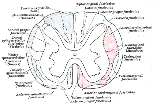 Diagram of the principal fasciculi of the spinal cord. (Spinotectal fasciculus labeled at bottom left.) | |
| Details | |
| Identifiers | |
| Latin | tractus spinotectalis |
| TA98 | A14.1.02.224 A14.1.04.141 |
| TA2 | 6107 |
| FMA | 73968 |
| Anatomical terminology | |
The spinotectal tract and/or spinomesencephalic tract (also spinotectal fibers, spinomesencephalic fibers,[1] spinotectal fasciculus, or spino-quadrigeminal system of Mott[2]) is a component of the ascending reticular activating system that is involved in processing of pain and visceral sensations.[3] The tract is involved in the processing of pain sensation, and reflex turning of the head and trunk in the direction of painful stimuli.[1] It arises in the spinal cord, and projects contralaterally to various structures of the midbrain.
Sources may distinguish between a distinct spinotectal tract which projects to the visual reflex system, and spinomesencephalic tract which projects to structures involved in pain processing.[1]
Anatomy
[edit]The ST/SM tract crosses over (decussates).[3]
Origin
[edit]The ST/SM tract arises in the laminae I and V of the posterior grey column of the spinal cord.[1] It arises in same region of the spinal cord grey matter as the spinothalamic tracts.[3]
Projections
[edit]Note: rightward arrows indicate subsequent functionally relevant "downstream" projections/pathways of some targets of the ST/SM tract.
- Periaqueductal gray → nucleus raphe magnus and gigantocellular reticular nucleus of the reticular formation of the medulla oblongata (enkephalinergic excitatory synapse) - modulates nociception through the serotonergic-opioid peptide descending pain-inhibiting system.[1]
- Nuclei of the reticular formation[3]
- Mesencephalic raphe nuclei - modulate nociception through the serotonergic-enkephalinergic opioid peptide descending pain-inhibiting system.[1]
- Parabrachial nucleus → amygdala (the latter being involved in processing emotional responses).[1]
- Superior colliculus → tectospinal tract (mediates reflex movement of the head and eyes in the direction of origin of a noxious sensation).[1]
- Pretectum[1]
See also
[edit]References
[edit]- ^ a b c d e f g h i Patestas, Maria A.; Gartner, Leslie P. (2016). A Textbook of Neuroanatomy (2nd ed.). Hoboken, New Jersey: Wiley-Blackwell. pp. 112, 203–204, 224–225. ISBN 978-1-118-67746-9.
- ^ Gray, Henry (1918). Gray's Anatomy (20th ed.). p. 762.
- ^ a b c d Kiernan, John A.; Rajakumar, Nagalingam (2013). Barr's The Human Nervous System: An Anatomical Viewpoint (10th ed.). Philadelphia: Wolters Kluwer Lippincott Williams & Wilkins. p. 74. ISBN 978-1-4511-7327-7.
External links
[edit]
This neuroanatomy article is a stub. You can help Wikipedia by expanding it. |