Endocast

An endocast is the internal cast of a hollow object, often specifically used for an endocasts of the cranial vault.[1] Endocasts can be man-made for examining the properties of a hollow, inaccessible space, or occur naturally through fossilisation.
Cranial endocasts
Manmade casts
Endocasts of the inside of the neurocranium (braincase) are often made in paleoanthropology to study brain structures and hemispheric specialization in extinct human ancestors. While an endocast can not directly reveal brain structure[citation needed], it can allow scientists to gauge the size of areas of the brain situated close to the surface, notably Wernicke's and Broca's areas, responsible for interpreting and producing speech.
Traditionally, the casting material is some form of rubber or rubber-like material. The openings to the brain cavity, except for the foramen magnum, are closed, and the liquid rubber inserted in the empty cranial vault to set. The resulting hollow sphere can then be drained of air like a balloon and pulled out through the foramen magnum.[2] Rubber endocasts like these were the standard practice until the end of the 20th century and are still used in some fields. However, scientists are increasingly utilizing computerized tomography scanning technology to create digital endocasts in order to avoid risking damage to valuable specimens.[3]
Natural endocasts
Natural cranial endocasts are also known. The famous Taung Child, the first Australopithecus found, consists of a natural endocast connected to the facial portion of the skull. It was the shape of the brain that allowed Raymond Dart to conclude that the fossil was that of a human relative rather than an extinct ape.[4]
Mammal endocasts are particularly useful as they resemble the fresh brain with the dura mater in place. Such "fossil brains" are known from several hundred different mammal species.[1] More than a hundred natural cast of the cranial vault of Bathygenys (a small merycodont) alone are known, some having identifiable features down to the major gyri.[5] A natural cranial endocast of a Tyrannosaurus brain vault is also known, showing the animal had limited intelligence, but a well-developed sense of smell.[6] The oldest known natural cranial endocast is a fossil fish brain from a Holocephalan, some 300 million years old.[7]
Endocasts of other hollows
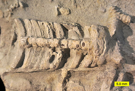
Endocasts fossils from animals with shells that easily disintegrate or dissolve, like the aragonite shells of certain molluscs and the tests of sea urchins can often be encountered free from their mold fossil. A frequent form is the internal mold of brachiopods. In the quite symmetrical genus Pentamerus the endocast resembles a vulva, giving these fossils the name Schamsteine ("shame stones") in German. The "Venus of Svinesund", a early mesolithic Venus figurine from Norway is a re-worked brachiopod endocast.[8] Endocasts are also known from snail shells and even from the stomach hollow of jellyfish, a group that rarely leave fossil traces.
Man-made endocasts are sometimes made from blood vessels for medical or anatomical reasons. The blood vessel of an organ (e.g. brain or liver) is injected with a resin. When it is set, the organ itself is dissolved, leaving a three-dimensional image of the blood supply to the organ.
References
- ^ a b Jerison, H.J. "Paleoneurology: The study of brain endocasts of extinct vertebrates". Comparative Mammalian Brain Collection. University of Wisconsin, Michigan State University, National Museum of Health and Medicine, sponsored by the National Science Foundation. Retrieved 17 November 2011.
- ^ McGowan, Christopher (1991). Dinosaurs, spitfires, and sea dragons (Compl. rev. and updated version of "The successful dragons" ed.). Cambridge, Mass.: Harvard University Press. ISBN 0-674-20769-6.
- ^ Brett-Surman, edited by M. K.; Buchholtz, E.; Jr., Thomas R. Holtz; director, James O. Farlow; Bob Walters, art. The complete dinosaur (2nd ed. ed.). Bloomington, Ind.: Indiana University Press. pp. 191–208. ISBN 978-0-253-00849-7.
{{cite book}}:|edition=has extra text (help);|first1=has generic name (help)CS1 maint: multiple names: authors list (link) - ^ Brain, C.K. Raymond Dart and our African Origins, in A Century of Nature: Twenty-One Discoveries that Changed Science and the World, Laura Garwin and Tim Lincoln, eds.
- ^ Wilson, J.A. (1971). "Early Tertiary vertebrate faunas, Vieja Group: Trans- Pecos Texas: Agriochoeridae and Merycoidodontidae". Texas Memorial Museum Bulletin (18): 1–83.
- ^ Australian museum: Tyrannosaurus rex brain
- ^ Oldest Fossil Brain Find Is 'Really Bizarre', LiveScience.com
- ^ Venus from Svinesund