Introduction
A modification of the basic immunofluorescent staining and flow cytometric analysis protocol can be used for the simultaneous analysis of surface molecules
and intracellular antigens at the single-cell level. In this protocol, cells are first activated in vitro, stained for surface antigens as in the surface antigen protocol, then fixed with paraformaldehyde to stabilize the cell membrane and permeabilized with the detergent saponin to allow anti-cytokine antibodies to
stain intracellularly. In vitro stimulation of cells is usually required for detection of cytokines by flow cytometry since cytokine levels are typically too low in
resting cells. Stimulation of cells with the appropriate reagent will depend on the cell type and the experimental conditions. For example, to stimulate T cells
to produce IFN-γ, TNF-α, IL-2, and IL-4, a combination of PMA (a phorbol ester / PKC activator) and Ionomycin (a calcium ionophore) or anti-CD3
antibodies can be used. To induce IL-6, IL-10 or TNF-α production by monocytes, stimulation of PBMC’s with lipopolysaccharide (LPS) can be used.
Note: Optimal stimulation period for induction of a given cytokine is variable and has to be determined. For example, the best time for detection of IL-6-producing cells by human
LPS-activated monocytes is 6 hours, whereas IL-10 detection needs at least 24 hours stimulation.
In contrast to detection of secreted cytokines by ELISA, for detection of intracellular cytokines, it is necessary to block secretion of cytokines with protein
transport inhibitors, such as Monensin or Brefeldin A, during the last few hours of the stimulation. It is advised that the investigators evaluate the use and efficacy of different protein transport inhibitors in their specific assay system.
Note: Generally, the non-specific background staining of fixed and permeabilized cells is higher than surface staining; therefore, extra protein such as BSA or FCS can be included in the staining buffer. The optimal concentration of the fluorochrome-conjugated anti-cytokine antibodies has to be determined experimentally. To confirm specificity of the staining, it is useful to block the directly-conjugated anti-cytokine antibodies with excess amounts of cognate ligand.
Buffers used for intracellular staining can have varying effects. eBioscience antibodies are optimized using eBioscience Foxp3 staining buffer set (cat. 00-5523) or eBioscience IC Fixation Buffer (cat. 00-8222). All antibodies to cytokines tested have stained appropriately using the Foxp3 buffers. Please contact Technical support for more information.
Granzyme B & IL-17 Data

|

|
|

|

|
Mouse/Human Granzyme B (GB11):
Left: Human peripheral blood cells were stimulated for 2 days with IL-2. The cells were surface stained with FITC CD56 (MEM188) (Cat. No. 11-0569) and stained intracellularly with PE GB11. Quadrants demarcate boundary for isotype controls.
Right: Mouse splenocytes were stimulated for 3 days with IL-2. Cells were surface stained with APC CD49d (DX5) (Cat. No. 17-5971) and subsequently stained intracellularly with PE GB11.
|
|
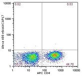
Human IL-17 (eBio64CAP17):
Human PBMCs were stimulated with TPA/ionomycin in the presence of monensin for 5 hours. The cells were fixed and permeabilized and intracellularly stained with anti-human CD4, clone RPA-T4, (Cat. No. 17-0049) and Alexa Fluor® 488 anti-human IL-17 (Cat. No. 53-7178). Isotype control on the left and anti-IL-17 on the right. Cells in the lymphocytes gate were used for analysis.
|
Intracellular Staining Quick Guides
Table 1: Mouse Cytokines: Intracellular Staining Quick Guide
|
Mouse Cytokines: Intracellular Staining Quick Guide
|
| Mouse Cytokine |
Cell Source |
Activation |
Incubation Time |
Restimulation |
Intracellular Block |
Antibody |
| GM-CSF
|
mouse spleen
|
ConA (3ug/ml) (2d)/IL-2 (20ng/ml)+IL-4 (20ng/ml) (3d)
|
2d/3d
|
anti-CD3 (10ug/ml immobilized) + anti-CD28 (2ug/ml soluble) 5hr
|
Brefeldin A
|
MP1-22E9
|
| IFN-γ
|
mouse spleen
|
ConA (3ug/ml) (2d)/IL-2 (20ng/ml)+IL-4 (20ng/ml) (3d)
|
2d/3d
|
anti-CD3 (10ug/ml immobilized) + anti-CD28 (2ug/ml soluble) 5hr
|
Brefeldin A
|
XMG1.2
|
| IL-1α
|
mouse PEC
|
mIFNγ (100ng/ml) (2hr)/LPS (100ng/ml)(22hr)
|
2hr/22hr
|
-
|
Brefeldin A
|
ALF-161
|
| IL-1β
|
mouse PEC
|
mIFNγ (100ng/ml) (2hr)/LPS (100ng/ml)(22hr)
|
2hr/22hr
|
-
|
Brefeldin A
|
polyclonal
|
| IL-2
|
mouse spleen
|
ConA (3ug/ml) (2d)/IL-2 (20ng/ml)+IL-4 (20ng/ml) (3d)
|
2d/3d
|
anti-CD3 (10ug/ml immobilized) + anti-CD28 (2ug/ml soluble) 5hr
|
Brefeldin A
|
JES6-5H4
|
| IL-4
|
mouse spleen
|
ConA (3ug/ml) (2d)/IL-2 (20ng/ml)+IL-4 (20ng/ml) (3d)
|
2d/3d
|
anti-CD3 (10ug/ml immobilized) + anti-CD28 (2ug/ml soluble) 5hr
|
Brefeldin A
|
BVD6-24G2,
11B11
|
| IL-5 NEW!
|
mouse splenic CD4
|
ConA (3ug/ml) (2d)/IL-2 (20ng/ml)+IL-4 (20ng/ml) (3d)
|
2d/3d
|
anti-CD3 (10ug/ml immobilized)+anti-CD28 (2ug/ml soluble) 5hr
|
Brefeldin A
|
TRFK5
|
| IL-6
|
mouse PEC
|
mIFNγ (100ng/ml) (2hr)/LPS (100ng/ml)(22hr)
|
2hr/22hr
|
-
|
Brefeldin A
|
MP5-20F3
|
| IL-10
|
mouse spleen
|
ConA (3ug/ml) (2d)/IL-2 (20ng/ml)+IL-4 (20ng/ml) (3d)
|
2d/3d
|
anti-CD3 (10ug/ml immobilized) + anti-CD28 (2ug/ml soluble) 5hr
|
Brefeldin A
|
JES5-16E3,
JES5-2A5
|
| IL-12/IL-23 (p40)
|
mouse PEC
|
mIFNγ (100ng/ml) (2hr)/LPS (100ng/ml) (22hr)
|
2hr/22hr
|
-
|
Brefeldin A
|
C17.8
|
| IL-17A NEW!
|
CD4+CD25- T cells from mouse spleen plus day 10 bone marrow-derived dendritic cells
|
anti-CD3, anti-IL-4, anti-IFN-γ, recTGF-β, recIL-6
|
4d
|
PMA/Iono 5hr
|
Monensin
|
eBio17B7
|
| IL-17F NEW!
|
mouse CD4
|
IL-17 induction
|
6d
|
PMA/Iono 5hr
|
Monensin
|
eBio18F10
|
| MCP-1/ CCL2 NEW!
|
mouse PEC
|
LPS 100ng/ml
|
24hr
|
-
|
Brefeldin A
|
2H5
|
| TNF-α
|
mouse PEC
|
mIFNγ (100ng/ml) (2hr)/LPS (100ng/ml)(22hr)
|
2hr/22hr
|
-
|
Brefeldin A
|
MP6-XT22,
TN3-19
|
| Annotations: mouse PEC=mouse thioglycolate-elicited peritoneal macrophages; ConA=Concanavalin A; Iono=Ionomycin; LPS=Lipopolysaccharide; PMA=Phorbol Myristate Acetate; 2d=2 day culture; 5hr=5 hour culture |
Table 2: Human Cytokines: Intracellular Staining Quick Guide
|
Human Cytokines: Intracellular Staining Quick Guide
|
| Human Cytokine |
Cell Source |
Activation |
Incubation Time |
Restimulation |
Intracellular Block |
Antibody |
| IFNα2
|
PMBC
|
CpG A ODN2216 (20ug/ml)
|
20 hours
|
-
|
Brefeldin A (added 2-4 hr post stimulation)
|
225.C
|
| IFN-γ
|
PBMC
|
PMA (30-50ng/ml)/Iono (1ug/ml)
|
5hr
|
-
|
Brefeldin A
|
4S.B3
|
| IL-1α
|
PBMC
|
LPS 100ng/ml
|
24hr
|
-
|
Brefeldin A
|
364/3B3-14
|
| IL-1β NEW!
|
PBMC
|
LPS 100ng/ml
|
24hr
|
-
|
Brefeldin A
|
CRM56
|
| IL-1RA
|
PBMC
|
LPS 100ng/ml
|
24hr
|
-
|
Brefeldin A
|
CRM17
|
| IL-2
|
PBMC
|
PMA (30-50ng/ml)/Iono (1ug/ml)
|
5hr
|
-
|
Brefeldin A
|
MQ1-17H12
|
| IL-4
|
PBMC
|
anti-CD3 (10µg/ml, immobilized) + anti-CD28 (2µg/ml, soluble) + IL-2 (10ng/ml) + IL-4 (20ng/ml) (2d); IL-2 (10ng/ml) + IL-4 (20ng/ml) (3d)
|
2d/3d
|
PMA (5ng/ml) + Ionomycin (500ng/ml) (4hr)
|
Brefeldin A
|
MP4-25D2,
8D4-8
|
| IL-5
|
CD4
|
anti-CD3 (10µg/ml, immobilized) + anti-CD28 (2µg/ml, soluble) + IL-2 (10ng/ml) + IL-4 (20ng/ml) (2d); IL-2 (10ng/ml) + IL-4 (20ng/ml) (3d)
|
2d/3d
|
PMA (5ng/ml) + Ionomycin (500ng/ml) (4hr)
|
Brefeldin A
|
TRFK5,
JES1-5A10
|
| IL-6
|
PBMC
|
LPS 100ng/ml
|
5hr
|
-
|
Brefeldin A
|
MQ2-13A5
|
| IL-10
|
PBMC
|
Th2 polarizing cultures
|
6d
|
PMA (50ng/ml) + ionomycin (1ug/ml)
|
Monensin
|
JES3-9D7
|
| IL-12/ IL-23 (p40)
|
PBMC
|
hIFNγ (100ng/ml) (2hr)/LPS (100ng/ml) (22hr)
|
2hr/22hr
|
-
|
Brefeldin A
|
C8.6
|
| IL-13 NEW!
|
CD4
|
anti-CD3 (10µg/ml, immobilized) + anti-CD28 (2µg/ml, soluble) + IL-2 (10ng/ml) + IL-4 (20ng/ml) (2d); IL-2 (10ng/ml) + IL-4 (20ng/ml) (3d)
|
2d/3d
|
PMA (5ng/ml) + Ionomycin (500ng/ml) (4hr)
|
Brefeldin A
|
PVM13-1
|
| IL-17A NEW!
|
PBMC
|
PMA (30-50ng/ml)/Iono (1ug/ml)
|
5hr
|
|
Monensin
|
eBio64CAP17,
eBio64DEC17
|
| IL-21
|
PBMC
|
PMA (30-50ng/ml)/Iono (1ug/ml)
|
4-7hr or 12-18hr
|
|
Brefeldin A
|
eBio3A3-N2
|
| MCP-1/ CCL2 NEW!
|
PBMC
|
LPS 100ng/ml
|
24hr
|
-
|
Brefeldin A
|
2H5,
5D3-F7
|
| TNF-α
|
PBMC
|
PMA (30-50ng/ml)/Iono (1ug/ml)
|
5hr
|
-
|
Brefeldin A
|
MAb11
|
| TNF-β
|
PBMC
|
anti-CD3 (10µg/ml, immobilized) + IL-2 (10ng/ml) (2d); IL-2 (10ng/ml) (3d)
|
2d/3d
|
PMA (5ng/ml) + Ionomycin (500ng/ml) (6hr)
|
Brefeldin A
|
359-81-11
|
| Annotations: Iono=Ionomycin; PMA=Phorbol Myristate Acetate; LPS=Lipopolysaccharide; 2d=2 day culture; 5hr=5 hour culture; LPS for activation of human PBMC obtained from Sigma (#L-8274) |