Ultrasound - Pelvis
|
Click images to view larger

More images
|
What is Pelvic Ultrasound Imaging?
Ultrasound imaging, also called ultrasound scanning or sonography, involves exposing part of the body to high-frequency sound waves to produce pictures of the inside of the body. Ultrasound exams do not use ionizing radiation (x-ray). Because ultrasound images are captured in real-time, they can show the structure and movement of the body's internal organs, as well as blood flowing through blood vessels.
Ultrasound imaging is usually a painless medical test that helps physicians diagnose and treat medical conditions.
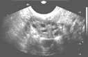
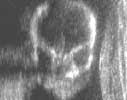
A pelvic ultrasound provides pictures of the structures and organs in the lower belly or pelvis.
There are three types of pelvic ultrasound:
A Doppler ultrasound exam may be part of a pelvic ultrasound examination.
Doppler ultrasound is a special ultrasound technique that evaluates blood as it flows through a blood vessel, including the body's major arteries and veins in the abdomen, arms, legs and neck.
What are some common uses of the procedure?
In men and women, a pelvic ultrasound exam can help identify:
Pelvic ultrasound is also used to guide procedures such as needle biopsies, in which needles are used to extract a sample of cells from organs for laboratory testing.
 In women, a pelvic or abdominal ultrasound is most often performed to evaluate the: In women, a pelvic or abdominal ultrasound is most often performed to evaluate the:
Pelvic ultrasound exams are also used to monitor the health and development of an embryo or fetus during pregnancy (see the Ultrasound-Obstetric page).
Ultrasound examinations can help diagnose symptoms experienced by women such as:
- pelvic pain
- abnormal bleeding
- other menstrual problems
and help identify:
A transvaginal ultrasound is usually performed to view the endometrium or the lining of the uterus, including its thickness and any associated ovarian abnormality. Transvaginal ultrasound also affords a good way to evaluate the muscular walls of the uterus, called the myometrium. Hysterosonography allows for a more in-depth investigation of uterine cavity. These exams are typically performed to detect:
- uterine anomalies
- scars
- polyps
- fibroids
- cancer, especially in patients with abnormal uterine bleeding
Some physicians also use hysterosonography for patients with infertility. See the Hysterosonography page for more information.
In men, a pelvic or abdominal ultrasound is used to evaluate the:
The transrectal ultrasound, a special study of the prostate gland, involves attaching the transducer to a probe and inserting it into a man's rectum. See the Prostate Ultrasound page for more information.
Doppler ultrasound images can help the physician to see and evaluate:
- blockages to blood flow (such as clots)
- narrowing of vessels (which may be caused by plaque)
- tumors and congenital malformation
How should I prepare for the procedure?
You should wear comfortable, loose-fitting clothing for your ultrasound exam. You will need to remove all clothing and jewelry in the area to be examined.
You may be asked to wear a gown during the procedure.
A full bladder helps to visualize the uterus, ovaries, bladder wall and prostate gland.
What does the equipment look like?
Ultrasound scanners consist of a console containing a computer and electronics, a video display screen and a transducer that is used to scan the body and veins. The transducer is a small hand-held device that resembles a microphone, attached to the scanner by a cord. The transducer sends out high frequency sound waves and then listens for the returning echo. The principles are similar to sonar used by boats and submarines.
The ultrasound image is immediately visible on a nearby screen that looks much like a computer or television monitor. The image is created based on the amplitude (strength), frequency and time it takes for the sound signal to return from the patient to the transducer.
For ultrasound procedures requiring insertion of the transducer, such as transvaginal or transrectal exams, the device is covered and lubricated.
How does the procedure work?
Ultrasound imaging is based on the same principles involved in the sonar used by bats, ships and fishermen. When a sound wave strikes an object, it bounces backward, or echoes. By measuring these echo waves it is possible to determine how far away the object is and its size, shape, consistency (whether the object is solid, filled with fluid, or both) and uniformity.
In medicine, ultrasound is used to detect changes in appearance and function of organs, tissues, or abnormal masses, such as tumors.
In an ultrasound examination, a transducer both sends the sound waves and records the echoing waves. When the transducer is pressed against the skin, it directs a stream of inaudible, high-frequency sound waves into the body. As the sound waves bounce off of internal organs, fluids and tissues, the sensitive microphone in the transducer records tiny changes in the sound's pitch and direction. These signature waves are instantly measured and displayed by a computer, which in turn creates a real-time picture on the monitor. These live images are usually recorded on videotape and one or more frames of the moving pictures are typically captured as still images.
The same principles apply to ultrasound procedures such as transrectal and transvaginal which require insertion of a special transducer into a natural opening in the body.
Doppler ultrasound, a special application of ultrasound, measures the direction and speed of blood cells as they move through vessels. The movement of blood cells causes a change in pitch of the reflected sound waves (Doppler effect). A computer collects and processes the sounds and creates graphs or pictures that represent the flow of blood through the blood vessels.
What will I experience during and after the procedure?
Most ultrasound examinations are painless, fast and easy.
For a transabdominal exam:
After you are positioned on the examination table, the radiologist or sonographer will spread some warm gel on your skin and then press the transducer firmly against your body, moving it back and forth over the area of interest until the desired images are captured. There may be varying degrees of discomfort from pressure as the transducer is pressed against the area being examined.
If scanning is performed over an area of tenderness, you may feel pressure or minor pain from the procedure.
Ultrasound exams in which the transducer is attached to probe and inserted into an opening of the body may produce minimal discomfort.
With transvaginal ultrasound, although the examination is often performed to look for a cause of pelvic pain, the sonogram itself should not be painful or significantly increase your discomfort. A vaginal sonogram is usually more comfortable than a manual gynecologic examination.
If no biopsy is required, transrectal ultrasound of the prostate is similar or may have less discomfort than a rectal exam performed by your doctor.
If a biopsy is performed, additional discomfort, due to the needle insertion, is usually minimal because the rectal wall is relatively insensitive in the region of the prostate. A biopsy will add time to the procedure.
If a Doppler ultrasound study is performed, you may actually hear pulse-like sounds that change in pitch as the blood flow is monitored and measured.
Once the imaging is complete, the gel will be wiped off your skin.
After an ultrasound exam, you should be able to resume your normal activities.
Who interprets the results and how do I get them?
A radiologist, a physician specifically trained to supervise and interpret radiology examinations, will analyze the images and send a signed report to your primary care or referring physician, who will share the results with you. In some cases the radiologist may discuss preliminary results with you at the conclusion of your examination.
What are the benefits vs. risks?
Benefits
- Ultrasound scanning is noninvasive (no needles or injections) and is usually painless.
- Ultrasound is widely available, easy-to-use and less expensive than other imaging methods.
- Ultrasound imaging uses no ionizing radiation.
- Ultrasound scanning gives a clear picture of soft tissues that do not show up well on x-ray images.
- Ultrasound causes no health problems and may be repeated as often as is necessary if medically indicated.
- Ultrasound is the preferred imaging modality for the diagnosis and monitoring of pregnant women and their unborn infants.
- Ultrasound provides real-time imaging, making it a good tool for guiding minimally invasive procedures such as needle biopsies and needle aspiration of fluid in joints or elsewhere.
- Pelvic ultrasound can help to identify and evaluate a variety of urinary and reproductive system disorders in both sexes without even the minimal risks associated with x-ray exposure.
Risks
What are the limitations of Pelvic Ultrasound Imaging?
Ultrasound waves are reflected by air or gas; therefore ultrasound is not an ideal imaging technique for the bowel. Barium exams and CT scanning are the methods of choice for bowel-related problems.
Ultrasound waves do not pass through air; therefore an evaluation of the stomach, small intestine and large intestine may be limited. Intestinal gas may also prevent visualization of deeper structures such as the pancreas and aorta. Patients who are obese are more difficult to image because tissue attenuates (weakens) the sound waves as they pass deeper into the body.
|